Treatment completed by Dr Pooneh Shahab
Full-Mouth Rehabilitation with Layered IPS e.max Restorations — Four-Year Follow-Up
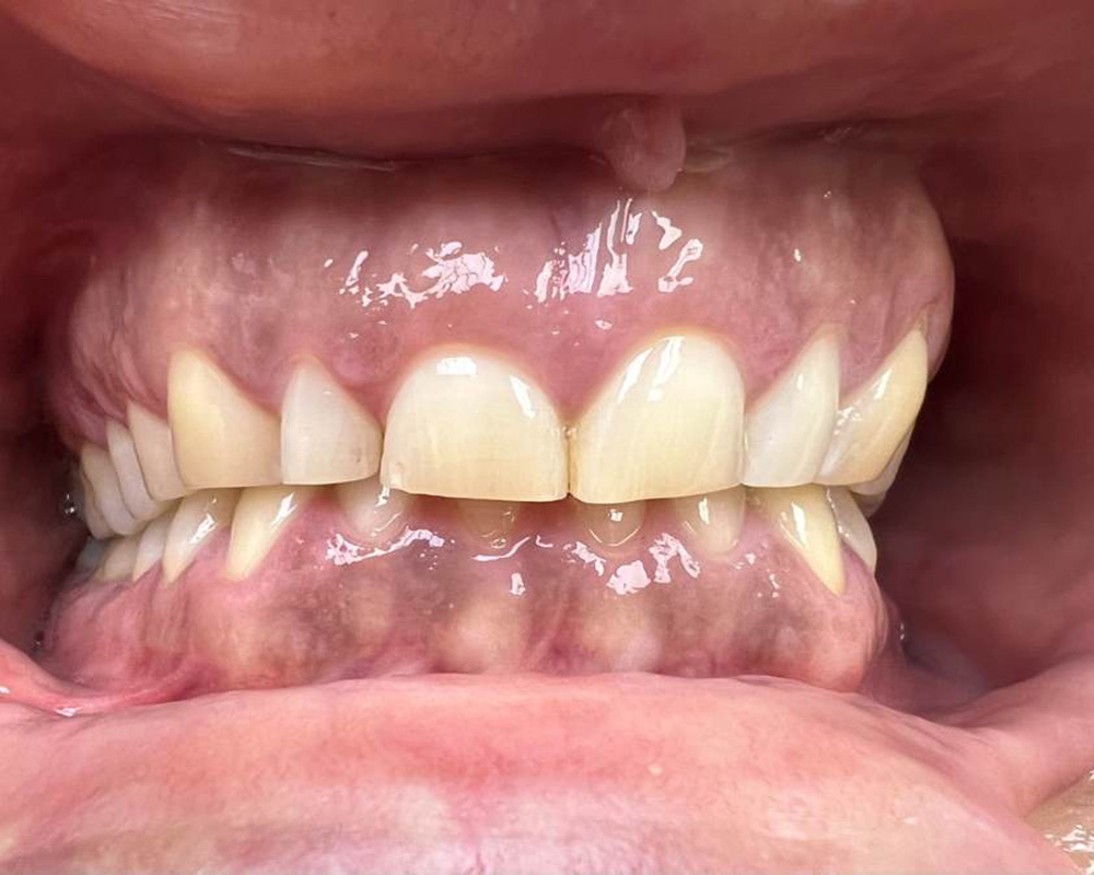 Initial consultation — Severe generalised tooth wear
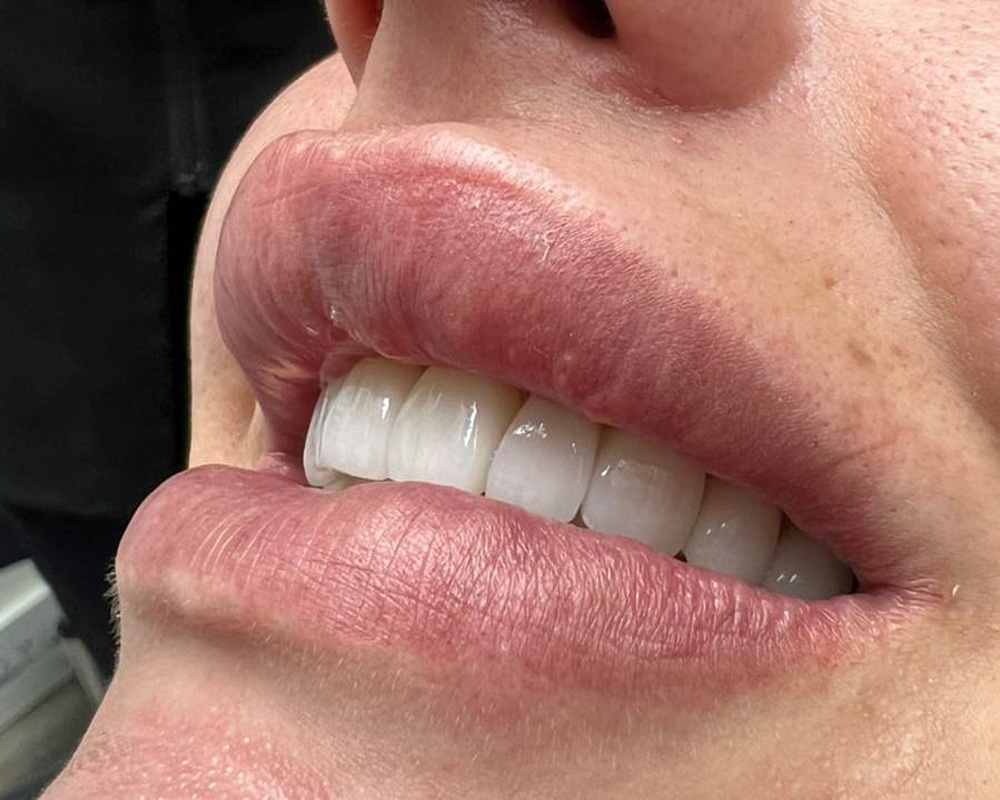 Four-year follow-up
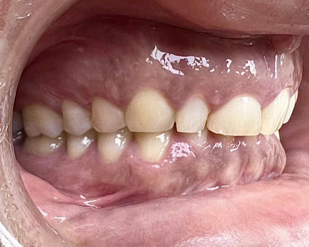 Initial consultation — Lateral intraoral view showing severe tooth wear
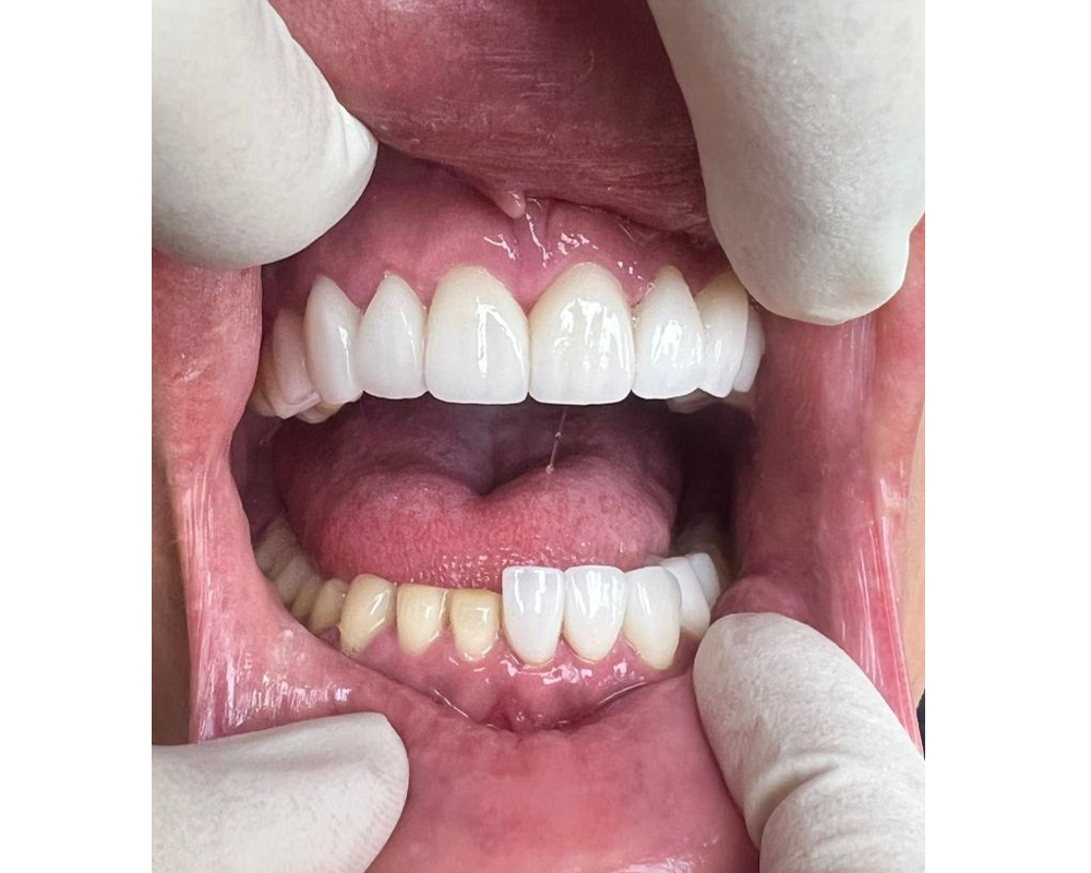 Veener delivery stage - Prepared teeth on the image left: veeners seated on the remaining teeth
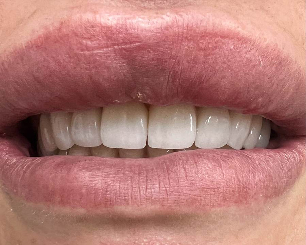 Four-year follow-up
Treatment delivery stage — The mandibular right teeth, visible on the left side of the photograph, are shown following conservative, individualised preparation and before delivery of their definitive restorations. This clinical view demonstrates the limited and tooth-specific preparation designs used during the rehabilitation.
Pre-treatment: Two photographs taken at the initial consultation demonstrate severe generalised tooth wear and substantial loss of natural tooth structure.
Treatment delivery: The delivery-stage photograph documents the conservative and individualised preparation designs used before the definitive restorations were delivered to the mandibular right teeth.
Post-treatment: Two photographs taken at the four-year follow-up demonstrate the frontal and oblique smile views.
This complex full-mouth rehabilitation addressed severe generalised tooth wear through a carefully planned, tooth-preserving restorative approach.
Laser reshaping of the gingival margins (laser gingivectomy) was performed where clinically indicated, increasing the visible clinical crown height by approximately 1 mm in selected areas.
Following detailed assessment of the patient’s bite, remaining tooth structure and available restorative space, the vertical dimension was carefully increased by approximately 3 mm anteriorly and 1.5 mm posteriorly. The bite was reorganised at a stable and reproducible jaw position.
The planned changes were assessed with provisional restorations for at least one month. During this phase, the patient’s comfort, function, adaptation and bite stability were reviewed before proceeding with definitive treatment.
Preparation design was individualised according to the condition and structural requirements of each tooth. Full circumferential crown preparation was not routinely used throughout the dentition. Instead, treatment incorporated a combination of veneers, partial-coverage and three-quarter preparations, and selected full-coverage crowns.
Palatal or lingual coverage was incorporated where required for function, restorative space and protection of the remaining tooth structure. This approach allowed healthy natural tooth structure to be preserved wherever clinically appropriate.
The delivery-stage photograph provides direct clinical documentation of this approach. The mandibular right teeth, visible on the left side of the photograph, had been conservatively and individually prepared, but their definitive restorations had not yet been delivered. The photograph therefore allows the limited extent and tooth-specific variation of the preparations to be observed.
All definitive ceramic restorations were fabricated using layered IPS e.max.
No teeth were extracted as part of the rehabilitation. One tooth required root canal treatment and was subsequently restored with a cast post and core before receiving its definitive layered IPS e.max crown. No other teeth required root canal treatment during the rehabilitation.
The assessment, treatment planning, laser gingivectomy, root canal treatment, cast post-and-core restoration, tooth preparations, provisional phase and delivery of the definitive restorations were completed by Dr Pooneh Shahab.
Important Information
Full-mouth rehabilitation is individually planned. Whether changes to tooth length, vertical dimension and bite are suitable and achievable depends on individual clinical findings, including the existing bite, remaining tooth structure, available restorative space, condition of the teeth and supporting tissues, smile line and adaptation during the provisional phase.
The type and extent of preparation required differ between individual teeth and patients. Veneers or partial-coverage restorations are not appropriate for every tooth, and full-coverage crowns may be required where clinically indicated.
Depending on the treatment required, potential risks may include sensitivity or pulpal complications that may require root canal treatment, gingival healing or contour changes, adaptation to changes in the bite, postoperative discomfort, loss of retention, chipping or fracture of ceramic restorations, complications affecting a root-canal-treated or post-retained tooth, and the need for ongoing maintenance, repair or replacement.
The outcomes shown are specific to this patient and do not necessarily reflect the results other patients may experience.